Basal cell carcinoma (BCC) is the most common type of skin cancer, with an estimated 4.3 million cases diagnosed per year in the United States. BCC results from the uncontrolled growth of cells from the outermost layer of the skin, the epidermis. BCC rarely spreads inside the body or travels into the bloodstream. Instead, it grows, infiltrates, and destroys the surrounding skin. Your dermatologist should treat BCC promptly in order to minimize the extent of infiltration into the surrounding skin and the resulting scarring from the treatment.
BCC does not evolve into malignant melanoma. Malignant melanoma is a different, more aggressive type of skin cancer. Individuals who have multiple BCCs or other skin cancers related to sun exposure are at an increased risk for malignant melanoma. However, if you have been diagnosed with BCC, you have a 50% chance of getting another one within the next 5 years. Therefore, it is important to follow up with your dermatologist every 6 months, or more often if appropriate, as well as to perform a skin self-exam weekly and to use sun protection daily.
BCC most often appears on sun-exposed areas, such as the face, scalp, ears, chest, back, arms, and legs of fair-skinned adults. BCC usually grows slowly over several months to years, but sometimes it grows quickly over weeks to months. Two common symptoms associated with BCC are itching and having a growth with a recurring cycle of bleeding and healing. The larger the BCC, the more complicated the treatment.
Risk factors
Anyone can get BCC, but there are certain risk factors that make some individuals more susceptible to BCC than others. Risk factors increase your susceptibility to BCC; however, they do not mean you will develop BCC.
Risk factors for BCC include
- Personal and family (genetic) history
- Fair skin with red hair and blue eyes
- Male over 50 years old
- Personal history of BCC, actinic keratosis, or squamous cell carcinoma
- Personal history of a rare genetic syndrome, such as basal cell nevus syndrome
- Environmental exposure
- Excessive long-term sun and ultraviolet light exposure
- Fair skin and having grown up in a southern region
- Frequent exposure to outside work or recreation
- History of multiple sunburns
- Freckles
- Use of an indoor tanning lamp or bed
- History of radiation therapy
- Medical condition that suppresses the immune system, such as AIDS or medications that organ transplant recipients take to suppress their immune system
- Excessive long-term sun and ultraviolet light exposure
What does BCC look like?
There are several types of BCC with different appearances:

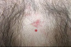
Figure 01

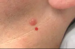
Figure 03
- Superficial BCC looks like a small, reddish, scaly patch of skin [Figure 1]
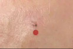
- Nodular BCC looks like a pearly, smooth, pimple-like lesion, almost translucent and skin colored [Figure 2]
- Pigmented BCC looks like a growth with brown and black tones often mimicking a malignant melanoma. Pigmented BCC is more common in darker skin types
- [Figure 3]
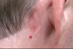
- Morpheaform / Infiltrating BCC looks like a smooth white scar, often difficult to diagnose, or like a growth or sore that slowly enlarges and keeps bleeding and healing over months [Figure 4]

Figure 02

Figure 04
How is BCC diagnosed?
Inspection of your skin by your dermatologist can confirm whether or not a growth is suspicious for BCC. If your dermatologist determines that a growth is suspicious for BCC then a biopsy will be performed. This is a simple procedure performed in the office under local anesthesia. Your growth will then be sent to a pathology lab where thin sections from the growth will be examined under a microscope by a dermatopathologist (a dermatologist or a pathologist trained in the microscopic examination of skin lesions). In the event your biopsy confirms BCC, your dermatologist will discuss treatment options.
Inspection of your skin at home with a weekly skin self-exam can help you identify a suspicious mole and help your dermatologist diagnose BCC early. [Table 1]
When inspecting your skin for any moles, growths, or spots, look for these signs.
| New | |
|---|---|
| Changing |
|
| Different and/or unusual |
A common symptom for BCC is a new growth with recurring cycles of bleeding and healing and itching. Be suspicious of any new spot or growth, changing spot or growth, or spot or growth that looks different or unusual from those in the surrounding area. If any spot or growth is suspicious, you should immediately report it to your dermatologist. Do not try to self-diagnose your condition.
Treatment options
There are many factors that can influence the choice of treatment:
- Type of BCC
- Location, size, number, and aggressiveness of BCC
- Patient’s general health
- Side effects, possible complications, benefits, and cure rate of a procedure
- Dermatologist’s experience and familiarity with a particular procedure
Each case is different. Your dermatologist will decide the most appropriate treatment plan for you. Commonly used procedures to treat AK include:
- Cryosurgery
- Topical chemotherapy
- Photodynamic therapy
- Electrodessication and curettage (ED&C)
- Surgical excision
- Mohs micrographic surgery
- Radiation therapy
For more information refer to “Common Procedures Performed in Dermatology”.
Follow-up care
Patients diagnosed with BCC should be examined by their dermatologist at least twice a year. Remember, 50% of individuals with a history of BCC will develop another one within 5 years of diagnosis. Your dermatologist will inspect your skin for any new BCCs and will ensure that any previously treated BCCs are not growing back.
Patients with a previous history of BCC should also perform a weekly skin self-exam. Learning what BCC looks like may help you identify a suspicious growth earlier. Inspecting the location of a previously treated BCC may also help you identify an early recurrence of BCC.
If you cannot see some part of your body, ask your partner or a family member to assist you with your weekly skin self-exam.


